Templates
Article.doc
Correspondance.doc
Evaluation ouverte.doc
Consentement eclaire.doc
Licence CC-BY-SA.doc

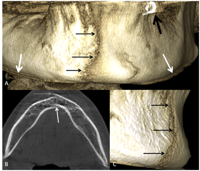
Stafne bone cavity (SBC) is a rare entity to find on panoramic radiography and on cone beam computed tomography. We reviewed in a systematic way the open-access literature from PubMed and DOAJ. We also proposed a new methodology consisting of collaboration with private practitioners, application of participative science approach, and open science practices, and using social media tool to obtain and describe seven different cases of SBC. We finally propose a new matrix table for classification of anatomical types of SBC already described and those yet to be described in open-access literature.

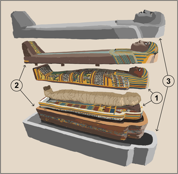
Objective: To perform a ‘virtual autopsy’ on the Egyptian mummy and to study, understand, and interpret three-dimensional (3D) high-resolution computed tomography (CT) scan images of Osirmose’s mummy with a multidisciplinary team composed of radiologists, archaeologists, and oral and maxillofacial surgeon.
Material and methods: We studied the Osirmose’s mummy, the doorkeeper of the Temple of Re, who lived during the XXVth dynasty. His mummy belongs to the Royal Museum of Art and History (Inv. E.5889). We performed a high resolution CT scanning of Osirmose’s mummy. We also 3D printed the upper maxilla of the mummy and a tooth found in the oesophagus with a clinically validated low-cost 3D printer.
Results: We confirmed the male sex of the mummy. We found the heart, aorta, and kidneys inside the mummy’s body. Brain excerebration was performed through the right ethmoid bone pathway. A wood stick embedded in the dura mater tissue was found inside the skull. The orbicularis oculi muscle, internal canthus, optical nerves, and calcified eye were still present. Artificial eyes were added above the stuffing of eye globes. The skull and face were embalmed with multiple layers of inner bandages in a sophisticated manner. The wear of maxillary teeth was asymmetrical and more pronounced on the maxilla. We discovered three anomalies of the upper maxilla: 1) a rectangular hole on the palatine side of tooth n°26 (the palatine root of tooth n°26 was missing), 2) an indentation at a right angle palatine to tooth n°27, and 3) a semilunar shape of edges around the osteolytic lesion distal and palatine to tooth n°28.
Conclusions: The present study provides the first evidence of a tooth removal site, and of oral surgery procedures previously conducted in a 2700-year-old Egyptian embalmed mummy. We found traces of dental root removal, and the opening of a tooth-related osteolytic lesion before the person’s death. The multidisciplinary team, the use of a high resolution 3D CT scan and a 3D-printed model of the upper maxilla helped in this discovery.
Les déplacements accidentels des dents de sagesse du maxillaire supérieur dans divers régions anatomiques sont rares. Nous avons effectué la recherche de littérature sur ce sujet de manière systématique en utilisant PubMed et DOAJ. Il n’existe pas d’illustration accessible gratuitement pour les voies de déplacements accidentels des dents de sagesse supérieurs imagées par le CT scan ou par le CBCT à part le déplacement vers la fosse infra-temporale et vers la fosse ptérygopalatine. Nous décrivons et illustrons par CBCT un cas unique dans la littérature médicale de déplacement accidentel du germe de la dent de sagesse du maxillaire supérieur dans l’espace jugal antérieur. Les raisons potentielles, les conséquences ainsi que les moyens de prévention de cette rare complication d’extraction de dents de sagesse sont aussi expliquées.

Objective: to know how much open access/open knowledge reference figures were available on motion artifacts in CBCT dentomaxillofacial imaging, and to describe and to categorize clinical variation of motion artifacts related to diverse types of head motion retrospectively observed during CBCT scanning time.
Material and methods: a search equation was performed on Pubmed database. We found 56 articles. The 45 articles were out of scope, and 7 articles were excluded after applying exclusion and inclusion criteria. Only 4 articles were finally freely accessible and selected for this review. Moreover, we retrospectively used our department CBCT database to search examinations with motion artifacts. We also checked retrospectively for radiological protocols as the type of motion artifact was described when occurred during the CBCT scanning time by the main observer. We had obtained the approval from the Ethical committee for this study.
Results: The accessibility of free figures on motion artifact in dentomaxillofacial CBCT is limited to 13 figures not annotated, and to one annotated figure presenting a double contour around cortex of bony orbits. We proposed to categorize the motion artifacts into three levels: low, intermediary, and major. Each level was related to: 1) progressive image quality degradation, 2) distortion of anatomy, and 3) potential possibility of performing clinical diagnosis. All 45 figures were annotated.
Conclusions: There exists a scarce open access literature on motion artifacts in CBCT. In our pictorial review we found that low level motion artifacts were more related to head rotation in axial plane (rolling). Rolling and lateral translation were responsible of intermediary level motion artifacts. Major level motion artifacts were created by complex motion with multiple rotation axes, multiple translation directions, and by anteroposterior translation. The main limitation of this study is related to retrospectively report empirical observation of patient motion during CBCT scanning and to compare these observations with motion artifacts found on clinical images. More robust methodology should be further developed using a virtual simulation of various types of head movements and associated parameters to consolidate the open knowledge on motion artifacts in dentomaxillofacial CBCT.

Objective: To investigate the participation of citizens-dental private practitioner in scientific articles about anatomical variations on dentomaxillofacial CBCT. Our null hypothesis was that private practice practitioners are not involved in publications on anatomical variations using cone beam computed tomography.
Material and methods: This study was performed from home without access to our university library. Only PubMed database was used to perform our study. We found 384 articles published among 1830 articles corresponding to our inclusion/exclusion criteria. For each selected article we searched for affiliation of all of the authors (university, private dental practice, students, other). We applied a co-creation approach to involve colleagues from private practice in analyzing results of this study.
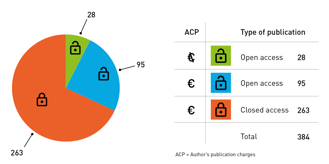
Results: A large majority of authors have university affiliation (96.5%). Only 3% of authors come from private practice. Most of articles belong to the group of 7 emergent economies (E7), and from Asia. 47.9% of 96 journals published only one article on anatomical variations discovered on CBCT. The higher number of articles (18.75%) were published by journals related to endodontics. The 84% of articles were dispersed among a vast span of general and specific dental, and maxillofacial journals. The 68.4% of articles on variations in CBCT were available in closed access and 31.6% of articles were available in open access. Only 6.7% of articles were published in open access without author publication charges (APC). The 31.6% of authors with university affiliation choose open access for their article. 7.8% of authors from private practice were involved in publishing in closed access journals and 2.34% in open access journals. Only 3 articles (0.78%) were published by authors affiliated to private practice without involvement of university authors. 2.6% of articles involved students as co-authors. Authors with other affiliation were involved only in one closed access publication. For the step of co-creation none of 183 private practitioners, and 3/33 (9%) university-affiliated members of Nemesis Facebook group actively participated in analyzing the results of this study.
Conclusions: the null hypothesis was accepted: dentists from private practice are exceptionally involved in publications on anatomical variations using CBCT in dentomaxillofacial area.
The objective of this work was to define the different criteria that a general dentist will have to take into account to equip himself with a three-dimensional (3D) printer for dental use. We have identified a total of 1037 3D printers produced by 342 companies and 211 3D printers from 88 companies that can print with 25µm layers. To be able to compare them, we evaluated 16 different characteristics: 1) family of 3D printing process, 2) minimum layer thickness, 3) presence or absence of scientific study to validate the minimum layer thickness, 4) minimal resolution on XY axes, 5) type of calibration, 6) printing environment, 7) presence of a heated printing plate, 8) maximum printing speed (in mm/s) with a link giving details of the layer thickness used, the XY resolution used and the material used to determine this speed, 9) dimensions of printing capacity, 10) capacity to use materials not originating from the manufacturer, 11) capacity to use biocompatible materials, 12) weight (in kg) and printer dimensions (in cm), 13) compatible operating systems, 14) compatible 3D print file types, 15) after-sales service and warranty period, 16) price, including whether taxes are included s or not. We noted a great heterogeneity of the information present, and information often absent regarding: 1) the type of calibration, 2) the printing speed, 3) the price, 4) the after-sales service, 5) the guarantee as well as 6) the materials which are taken into account by the 3D printer. We described multiple communication difficulties with our contacts and a very dynamic development of the 3D printing world. Finally, we proposed the characteristics of an "ideal" dental 3D printer and of an "ideal" partner company for a dentist wishing to obtain the 3D printer of his choice.
Cette revue illustrée porte sur les principales indications actuellement recommandées dans la littérature d’utilisation du cone beam computed tomography (CBCT) en orthodontie. Il s’agit des anomalies dentaires, des canines incluses, des dents surnuméraires, des troubles de l’éruption et des résorptions radiculaires externes liées aux traitements orthodontiques. L’examen CBCT doit être justifié individuellement, au cas par cas, et de pouvoir apporter un bénéfice au patient en terme de diagnostic et/ou de traitement orthodontique. L’orthodontiste prescripteur doit être capable d’interpréter et est responsable de l’interprétation de tout ce qui est visible sur l’ensemble du champs de vue du CBCT.
Objective: to explain the meaning and to illustrate technical artifacts (aliasing as well as the ring artifact) and beam hardening (metal artifact) that can be present in the dentomaxillofacial cone beam computed tomography (CBCT), and to check the accessibility of free illustrations of these artifacts in medical publications.
Material and methods: One observer applied five search equations using database PubMed. The exclusion criteria were: experimental studies, animal studies, studies not related to dentomaxillofacial area, and articles with closed access. There was no time limit for the search of articles. We limited our search to English and French language.
Results: Only 3 articles out of 434 publications were retained after application of inclusion/exclusion criteria. In these articles only 4 annotated figures were freely accessible in medical publications from PubMed.
In this paper we presented examples of aliasing, ring artifact, and beam artifacts from I-CAT, Carestream 9000 3D (Kodak), and Planmeca Promax 3D Mid CBCT. The intensity of beam hardening artifact varies from major degradation of image (i.e., subperiosteal implants, bridges, crowns, dental implants, and orthodontic fix appliances), through mean degradation (screws securing titanium mesh, head of mini-implant) to no beam hardening on metallic devices (orthodontic anchorage, orthodontic contention wire) or on dense objects (endodontic treatments, impression materials, Lego box). Some beam hardening artifacts arising from nasal piercing, hairs, or hearing aid device may be present on the image but they will not disturb the evaluation of the field of view.
Conclusions: reduction of aliasing artifact is related with the improvement of detectors quality. The presence of ring artifact means that CBCT device has lost its calibration. The field of view (FOV) needs to be reduce in order to avoid scanning regions susceptible to beam hardening (e.g., metallic restorations, dental implants). Finally, the accessibility to open knowledge on technique -related CBCT artifacts seems extremely limited when searching at PubMed database.
Objective: to investigate the accessibility of open access article on anatomical variations described on cone beam computed tomography (CBCT) using PubMed database. We wanted to investigate how many journals are sharing articles without pay-wall and how many are sharing articles without author publication charges.
Material and methods: a search equation was designed with exclusion criteria limiting the search in PubMed to articles published in English and French. The search was performed by one observer. We had found 2228 articles; among them 709 were accessible as ‘full text’. After applying exclusion criteria and after full text reading only 50 articles remained for the review.
Results: the 50 selected articles shared 306 annotated (visual marking, explanation like arrows) and 432 not annotated figures with the public. The 76% of articles were single studies on one specific topic. The main topic was endodontics with 22 articles. 28 journals from all continents participated in the effort of sharing of figures on anatomical variations from CBCT. However, only 2 journals were completely free of charges for authors and readers.
Conclusions: we have found only 15 annotated and 3 not annotated figures in 2 articles published in 2 different open access journals (without reader pay-wall and without author publication charges). Sharing the knowledge on anatomical variations from dentomaxillofacial CBCT represents an exception in dental literature.
Objective: To summarize the current knowledge on CT scanning of Egyptian mummy heads and faces and provide more valid methodology than that previously available.
Material and methods: A systematic review was performed by one observer using two biomedical databases: PubMed and EMBASE. Inclusion and exclusion criteria were applied along with language restrictions. Finally, 2120 articles were found, 359 articles were duplicated among all search equations, 1454 articles were excluded, 307 articles were retained for full review, and 28 articles (31 mummies) were selected for the final study (PRISMA workflow).
Results: The data were categorized into the following groups: 1) general information; 2) 1st author affiliation; 3) CT radiological protocol; 4) excerebration pathways; 5) soft tissue preservation; 6) dental status and displaced teeth; 7) packing of the mouth, ears, nose, and eyes, and 8) outer facial appearance. The evidence-based quality of the studies was low because only case reports and small case series were found.
Discussion: The embalming art applied to a mummified head and face shows great variability across the whole span of Egyptian civilization. The differences among the various embalming techniques rely on multiple tiny details that are revealed by meticulous analysis of CT scans by a multidisciplinary team of experts.
Conclusion: There is a need for more systematization of the CT radiological protocol and the description of Egyptian mumm’y heads and faces to better understand the details of embalming methods.
Our aim was to perform a systematic open-access review of various complications reported for surgically assisted rapid maxillary expansion (SARME) procedures. There were 37 articles found in Pubmed using the search equation. Twelve articles were initially excluded according to the exclusion criteria. The 25 remaining articles were read in full for their descriptions of complications related to the SARME procedure in mature patients. The main reversible complications of SARME were infection, postoperative pain, and bleeding. There were also complications related to distractors, to secondary surgeries, and pterygomaxillary junction. The main non-reversible complications of SARME were associated with teeth, periodontal bone loss, and skull base fractures. Large field-of-view cone beam computed tomography (maxilla and skull base) should be implemented as initial planning tool to prevent many potential complications. The current trend for “minimally invasive” surgery in SARME might be, from an ethical point of view, transformed onto “minimally complicated” surgery as complication is still more harmful for any given patient than any potential perioperative surgical invasiveness.
La fibrodysplasia ossificans progressiva ou myosite ossifiante est une maladie autosomique dominante très rare. La maladie est caractérisée par l’apparition progressive de calcifications ectopiques dans les tendons, les muscles striés, les ligaments et les fascias. Elles entrainent une perte de mobilité progressive du corps allant jusqu’à un handicap physique sévère. Le présent travail est une revue systématique de la littérature concernant les aspects oro-faciaux de cette pathologie rare. Des pistes de prise en charge de ces patients au niveau dentaire et maxillo-facial sont aussi proposées.
La myosite ossifiante traumatique (MOT) du muscle temporal est une pathologie extrêmement rare. Une limitation de l’ouverture buccale est le symptôme clé. Les examens d’imagerie sont très utiles au diagnostic, le CT-scanner de la face étant l’examen le plus performant pour la détection de la MOT. Le meilleur traitement est la résection chirurgicale complète. Les cas de myosite ossifiante traumatique dans la région de la tête et du cou sont rares. Les atteintes au niveau du muscle temporal sont encore plus rares. Nous présentons ici le cas d’une ossification isolée du muscle temporal gauche après un polytraumatisme. L’intérêt de l’observation clinique du cas présenté est donc de documenter la littérature. Le diagnostic différentiel et les signes pathognomoniques présents sur les examens d’imagerie seront également détaillés.
Objective: to develop and test inter-observer reproducibility of instructions for authors quality rating (IAQR) tool measuring the quality of instructions for authors at journal level for a possible improvement of editorial guidelines.
Material and methods: instructions for authors of 75 dental and maxillofacial surgery journals were assessed by two independent observers using assessment tool inspired from AGREE with 16 questions and 1 to 4 points scale per answer. Two observers evaluated the instructions of authors independently and blind to impact factor of a given journal. Scores obtained from our tool were compared
with “journal impact factor 2013”.
Results: IAQR presented with an excellent interobserver reproducibility (κ= 0.81) despite a difference in data distribution between observers. There existed a weak positive correlation between IAQR and “journal impact factor 2013”.
Conclusions: The IAQR is a reproducible quality assessment tool at the journal level. The IAQR assess the quality of instruction for authors and it is a good starting point for possible improvements of the instructions for authors, especially when it comes to their completeness.
Nemesis relevance: 28% of dental and maxillofacial journals might revise their instructions for authors to provide more up-to-date version.
Objective: Paget’s disease of bone is characterized by a focal increase in bone
resorption and accelerated bone formation leading to a weaker and disorganised bone. Bisphosphonates (BPs) have been the treatment of choice of Paget’s disease since the 1990s. Medication related osteonecrosis of the jaw (MRONJ) is a rare event in non oncologic patients. We describe a rare case of Paget’s disease
involving the maxilla with osteonecrosis in a context of bisphosphonate treatment.
Case report: an 87-year-old woman presented with 4 episodes of bone necrosis in 15 years. In this case report there is a clear chronologic association between the occurrence of MRONJ and the administration of iv BP for Paget’s disease.
Maxillofacial involvement of Paget’s disease occurs in less than 15% of cases. There is a lack of information in the literature about the association of MRONJ and Paget’s disease. Even if osteonecrosis of the jaw could be a consequence of the
disease, in this case, it is more in relation to the BP treatment.
Conclusions: Although MRONJ might be considered a rare condition in Paget’s disease, patients prior to starting antiresorptive therapy and in particular iv BPs should have a complete dental examination and panoramic X-Ray.
Nemesis relevance: side effect of bisphosphonate treatment

Objective: Our study aimed to determine the possibility of using models created with a low-cost, paper based 3D printer in an operating room. Therefore influence of different methods of sterilization on models was tested and cytotoxicity of generated models was determined.
Material and methods: 30 cuboids divided into three groups were used for verification of shape stability after sterilization. Each group was sterilized either with: Ethylene oxide in temperature 55˚C, Hydrogen peroxide gas plasma in temperature 60˚C or Gamma irradiation at 21˚C, 25kGy. Each cuboid was measured using calliper three times before and three times after sterilization. Results were analysed statistically in Statgraphics Plus. Statistical significance was determined as p< 0.05. Sixty cylinders divided into six groups were used for cytotoxicity tests. Three of those groups were covered before sterilization with 2-octyl-cyanoacrylate. Each group was sterilized with one of the previously described methods. Cytotoxicity was tested by Nanostructural and Molecular Biophysics Laboratory in Technopark Lodz using normal adult human dermal fibroblasts. Survival of cells was tested using spectrophotometry with XTT and was defined as ratio of absorbency of tested probe to absorbency of control probe. Calcein/Ethidium dyeing test was performed according to LIVE/DEAD Viability/Cytotoxicity Kit protocol. Observation was done under Olympus GX71 fluorescence microscope. Results: There was no statistically significant difference for established statistical significance p=0.05 in cuboids dimensions before and after sterilization regardless of sterilization method. In XTT analysis all samples showed higher cytotoxicity against normal, human, adult dermal fibroblast culture when compared to positive control. ANOVA statistical analysis confirmed that 2-octyl cyanoacrylate coating of paper model improved biological behaviour of the material. It decreased cytotoxicity of the model independently of sterilization method. In calcein/ethidium dyeing test due to the high fluorescence of the background caused by cylinders of analysed substance it was impossible to perform the exact analysis of the number of marked cells.
Conclusions: Acquired results allow to conclude that Mcor Technology Matrix 300 3D paper-based models can be used in operating room only if covered with cyanoacrylate tissue adhesive.
Nemesis relevance: no statistically significant difference in cuboids dimensions before and after sterilization regardless of sterilization method. Presence of high cytotoxicity of 3D paper-based models without coating.
Objectives: The Pierre Robin sequence (PRS) is defined by retromicrognathia, glossoptosis, and sleep apnea and can also be associated with cleft palate. Diagnosis, management and mandibular catch-up growth are still controversial issues in PRS patients. The aim of our retrospective study was to evaluate in three dimensions (3D) the airway space and mandibular morphology in PRS compared to a normal control group patients in the pre-orthodontic period of life. The null hypothesis was that we would not find a significant difference between the PRS and control group patients in oropharyngeal airway volume measurements. Material and methods: We analyzed 9 PRS patients (mean age: 8 years-old) who underwent cleft palate surgery in the first four months of life, performed by the same surgeon using the same technique. Cone-beam computed tomography (CBCT) was performed in these patients after local ethical committee approval. The control group consisted of 15 patients (mean age: 9 years-old) with CBCT already performed for other reasons. 3D Slicer was used in both groups for semi-automatic segmentation of the airway space. Two independent observers performed semi-automatic segmentations twice in each patient with a one- week interval between the two series of measurements. Airway volume was automatically measured using 3D Slicer. We also developed a 3D cephalometric analysis with Maxilim software in order to define a 3D mandibular morphology which consisted of 25 landmarks, 4 planes, and 23 distances. Two independent observers performed the 3D cephalometric analysis twice for each patient, with a one- week interval between the two series of measurements. Results: There was no significant difference in the intra- and inter-observer measurements between the PRS and control groups for airway space volume (p<0.05). However, there was a significant difference in the shape of the mandible between the PRS group and the control group (p<0.05). Conclusions: Vertical ramus width and mandibular global anteroposterior length were significantly lower in the PRS group. Mandibular hypoplasia could be found in PRS patients not only in the horizontal dimension. Nemesis relevance: the null hypothesis was confirmed. Moreover we failed to find exactly the same control group under 9 years-old due to radioprotection restrictions of application of cone beam CT in children.
Plusieurs pathologies osseuses des maxillaires ont comme caractéristique une tendance à la récidive, et ce même après un traitement approprié. Il convient donc de les reconnaître afin de pouvoir les surveiller, à la fois cliniquement et par une technique d’imagerie appropriée. On distinguera ici des pathologies tumorales bénignes et malignes, métaboliques, malformatives et infectieuses. Actuellement, le CT scan est la technique d’imagerie médicale de premier choix pour établir le diagnostic différentiel et permettre la surveillance de ces pathologies osseuses. Le traitement de ces pathologies récidivantes est soit le curetage/énucléation si possible (corticales non rompues) soit une résection interruptrice ou non des maxillaires ou de la mandibule, et ce en fonction des données cliniques et surtout anatomopathologiques à discuter et envisager au cas par cas, la généralisation dans ce domaine étant impossible voire dangereuse pour le patient. Il ne peut donc être question de proposer des recommandations ni des algorithmes décisionnels. Mots clés: pathologies osseuses, récidive, maxillaire, mandibule


