Dental use of cone beam computed tomography in pediatric embolized arteriovenous maxillofacial malformation
DOI :
https://doi.org/10.14428/nemesis.v20i1.63163Mots-clés :
pediatric, arteriovenous malformation, CBCT, embolization, cone beam computed tomographyRésumé
Objective: Pediatric facial arteriovenous malformations (AVMs) are rare but can cause potentially fatal hemorrhages during dental procedures and oral surgery. In this article we present a systematic review of the medical open access literature on pediatric facial AVM.
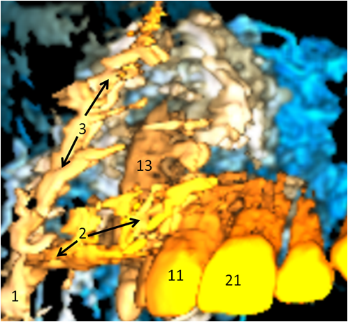
Case report: We illustrate our purpose with clinical dental use of cone beam computed tomography (CBCT) in pediatric embolized facial AVM to define the presence and the position of the right upper impacted canine.
Conclusions: We advocate the use of CBCT as additional imaging tool in the follow-up of pediatric dentomaxillofacial AVM, and for depiction of dentoalveolar structures that are inaccessible by conventional dental radiography.
Téléchargements
Publiée
Numéro
Rubrique
Licence
(c) Tous droits réservés Raphael Olszewski, Stéphanie Theys 2021

Ce travail est disponible sous licence Creative Commons Attribution - Partage dans les Mêmes Conditions 4.0 International.


