Roadmap for daily practice of CBCT in cleft lip palate paediatric patients: a pictorial review.
DOI:
https://doi.org/10.14428/nemesis.v30i1.77213Keywords:
cone beam computed tomography, CBCT, cleft lip palate, paediatric, reportingAbstract
Objective: to present and to illustrate a new methodology for daily practice in cone beam computed tomography (CBCT) interpretation and reporting in cleft lip palate (CLP) non syndromic paediatric patients. The proposed protocol is based on clinical experience and on systematic search of the literature.
Material and methods: We performed two types of systematic search of articles: 1) articles related to the use of CBCT in CLP patients, and 2) articles related to the reporting and interpretation of the CBCT images by radiologists. We used two databases PubMed and Google scholar.
Results: For indications of CBCT in CLP patients we found in PubMed 378 articles and 48 articles were selected for the review; in Google scholar we found 463 articles, and 9 articles were selected for the review. 2) For reporting in CBCT we found 956 articles in PubMed, and 9 articles were selected for the review.
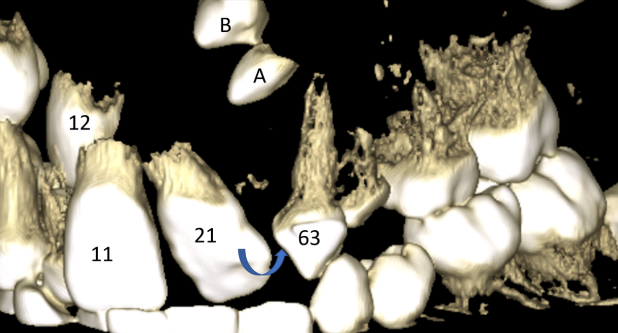
Conclusions: We presented the 6-steps system for interpretation and reporting information from CBCT of CLP paediatric patients: 1) Step 1 (axial view): presence or absence of bone bridge remnants of alveolar bone graft; Step 2 (3D dental tissue reconstruction): description of dental arch tooth by tooth, search for agenesis and supernumerary teeth, description of variation in the position of the tooth explaining the type of existing translation and rotation; Step 3 (coronal view): cleft palate pathway and its extension; anomaly in maxillary, ethmoid and sphenoid sinuses if existing; Step 4 (sagittal and coronal view): checking of the opening (calcification sites) of the sphenooccipital synchondrosis, and checking of anomalies of the occipital bone; Step 5 (3D bone tissue reconstruction): C1-C2 vertebra anomalies; Step 6 (3D soft tissue reconstruction): external ear anomalies. We illustrated our methodology with 46 figures from 5 CBCT of CLP patients.
Downloads
Published
Issue
Section
License
Copyright (c) 2023 Raphael Olszewski, Antoine De Muylder, Sergio Siciliano

This work is licensed under a Creative Commons Attribution-ShareAlike 4.0 International License.


