Diagnostic value of cone beam computed tomography in complex and compound odontomas: a systematic review and open classification matrix
DOI:
https://doi.org/10.14428/nemesis.v23i1.65993Keywords:
cone beam computed tomography, CBCT, complex odontoma, compound odontoma, odontogenic tumoursAbstract
Objective: Firstly, this review aims to analyse the recent literature about three-dimensional (3D) diagnostic imaging in complex and compound odontomas and compare it to two-dimensional (2D) imaging. Panoramic radiographs help to
evaluate the vertical position of odontomas, and occlusal radiographs are used to evaluate the proximity to adjacent teeth. However, cone beam computed tomography (CBCT) can offer volumetric images, and therefore, a more accurate three-dimensional analysis. Secondly, this research aims to construct an open classification matrix for complex and compound odontomas for dentomaxillofacial CBCT radiology protocols based on a systematic literature review.
Material and methods: Two systematic literature searches were conducted in PubMed (Medline), on 2 February 2022 concerning classification systems, and on 5 February 2022 concerning CBCT images.
Results: In total, these searches revealed 391 papers by reviewing the databases mentioned above. Six articles were selected for inclusion on classification of odontomas and 13 articles were found on CBCT imaging. Consequently, the
construction of an open classification matrix for compound and complex odontomas for dentomaxillofacial CBCT radiology protocols was performed using these 19 articles.
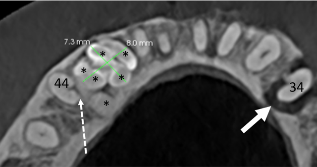
Conclusions: CBCT offers a more precise position and accurate diagnosis of complex and compound odontomas compared to 2D imaging. Consequently, it enhances the detailed view of the site (multiple or unique), location (intraosseous, partially or completely extragnathic), size, extension (bony expansion, thinning or perforation cortical bone), density and type (denticulo type, particle type, denticulo-particle type, denticulo-amorphous type, amorphous tissue), relationship (with the crown or root of the definitive tooth), adjacent teeth resorption (deciduous or definitive), adjacent teeth (retention or impaction), and distance with adjacent structures (inferior alveolar nerve, sinus maxillaris), as well as adequate surgical planning. Moreover, this research presents an open classification matrix for the most complete description of compound and complex odontomas when analysing CBCT imaging.
Downloads
Published
Issue
Section
License
Copyright (c) 2022 Kathia Dubron, Anna Gurniak, Eliza Gurniak, Constantinus Politis, Raphael Olszewski

This work is licensed under a Creative Commons Attribution-ShareAlike 4.0 International License.


